42 label this transmission electron micrograph
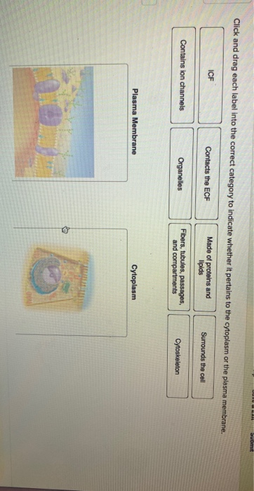
Label This Transmission Electron Micrograph / Microscopy Innovations ... Label This Transmission Electron Micrograph / Microscopy Innovations Transmission Electron Microscopy Tem - Abbie Bryan To leave a comment, click the button below to sign in with Google. Sign in with Google Join Our Newsletter Lap Practical #1 EC Flashcards | Quizlet Place the following cytoplasmic structures in the appropriate structural category. Label the transmission electron micrograph of the cell. What would be the consequence if the highlighted structures suddenly became nonpolar? The lipid bilayer would not be able to hold its shape in water and the cell membrane would disassemble.
The Transmission Electron Microscope | CCBER - UC Santa Barbara What is a Transmission Electron Microscope? Transmission electron microscopes (TEM) are microscopes that use a particle beam of electrons to visualize specimens and generate a highly-magnified image. TEMs can magnify objects up to 2 million times. In order to get a better idea of just how small that is, think of how small a cell is.

Label this transmission electron micrograph
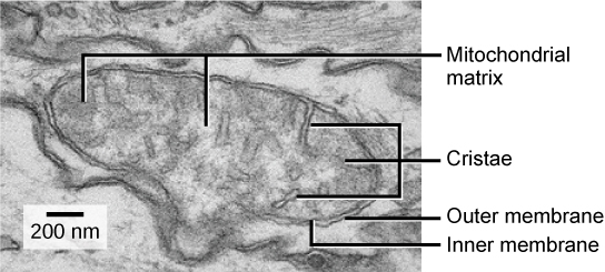
Solved Label the transmission electron micrograph of the | Chegg.com Expert Answer. Answer The label is indicated from TOP to BOTTOM Cil …. Label the transmission electron micrograph of the cilium. Microvillus Axoneme Cilium Dynein arm. Labeling the Cell Flashcards | Quizlet Label the transmission electron micrograph of the nucleus. membrane bound organelles golgi apparatus, mitochondrion, lysosome, peroxisome, rough endoplasmic reticulum nonmembrane bound organelles ribosomes, centrosome, proteasomes cytoskeleton includes microfilaments, intermediate filaments, microtubules Identify the highlighted structures Solved Label the transmission electron micrograph of the - Chegg Label the transmission electron micrograph of the mitochondrion. Matrix granule Mitochondrion Outer membrane Cristae Inner membrane Matrix Reset Zoom Question: Label the transmission electron micrograph of the mitochondrion. Matrix granule Mitochondrion Outer membrane Cristae Inner membrane Matrix Reset Zoom This problem has been solved!
Label this transmission electron micrograph. Label the transmission electron micrograph of the nucleus. - Transtutors Label the transmission electron micrograph of the nucleus. The Florida lottery has a game called Pick 4, where a player pays $1 and then picks a sequence of four numbers 0-9 (e.g., 1234, 0389, 9020). CH 9 skeletal muscle Flashcards | Chegg.com Label this transmission electron micrograph of relaxed sarcomeres by clicking and dragging the labels to the correct location. The electrical properties of cells are the result of Ion concentration differences across the plasma membrane. Check all that apply as it pertains to functions of the muscular system. 1. Transmission Electron Microscope (TEM) micrograph | Chegg.com 1. Transmission Electron Microscope (TEM) micrograph of Human Liver Cell from 8000X to 16000X 2. Scanning Electron Microscope (SEM) micrograph of COVID-19 Coronavirus from 100,000X to 200,000X 3. Light Microscope (LM) micrograph of MRSA (Methicillin Resistant Staphylococcus Aureus) from 400X to 1000X 4. Label This Transmission Electron Micrograph - Kaiden Brown Label This Transmission Electron Micrograph : TEM of chloroplast from Coleus blumei - Stock Image - B110 - Kaiden Brown To leave a comment, click the button below to sign in with Google. Sign in with Google Join Our Newsletter
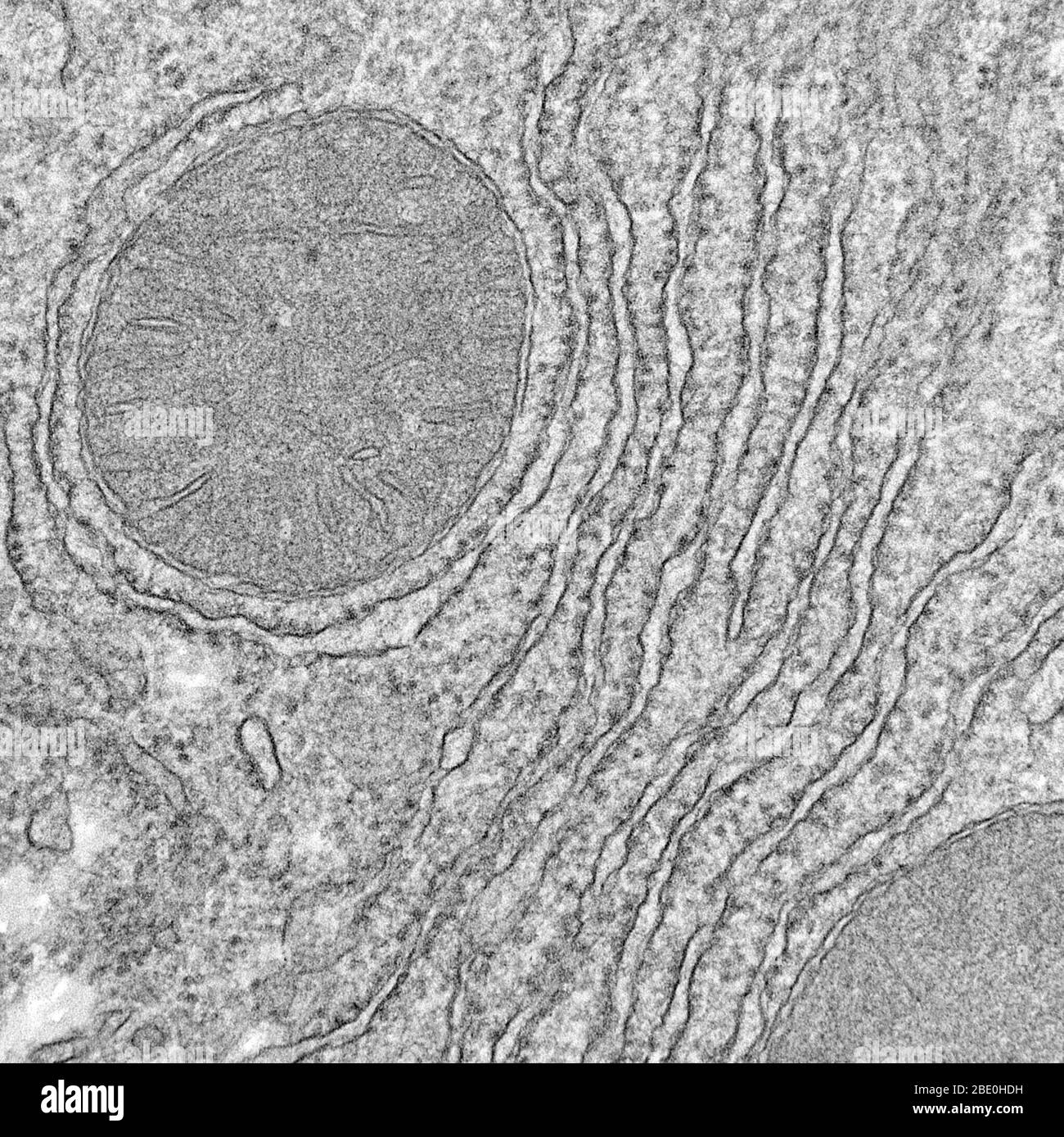
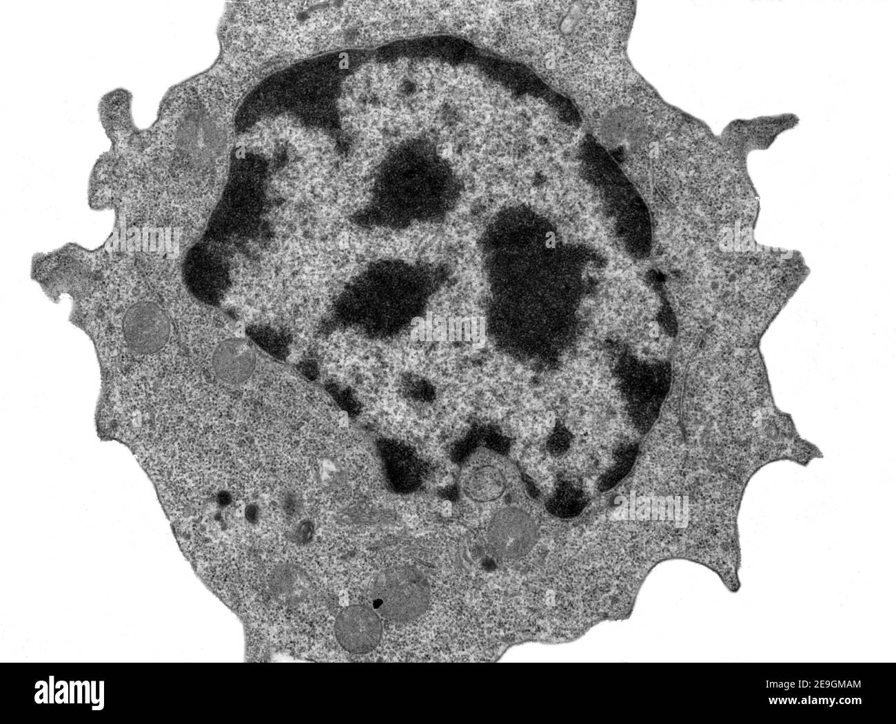
Solved Label the transmission electron micrograph of the - Chegg Question: Label the transmission electron micrograph of the nucleus. Nuclear envelope Nucleolus Nucleus Heterochromatin Reset Zoom This problem has been solved! You'll get a detailed solution from a subject matter expert that helps you learn core concepts. See Answer Show transcribed image text Expert Answer 100% (24 ratings) (Solved) - Label this transmission electron micrograph of relaxed ... Label this transmission electron micrograph of relaxed sarcomeres by clicking and dragging the labels to the correct location Sarcamere 1 band (light) Z disc Mline Aband (dark) H zone Jul 24 2022 01:02 PM 1 Approved Answer Hitesh M answered on July 26, 2022 5 Ratings ( 17 Votes) Next Previous Related Questions Q: Solved Label this transmission electron micrograph of - Chegg Label this transmission electron micrograph of relaxed sarcomeres by clicking and dragging the labels to the correct location Sarcamere 1 band (light) Z disc Mline Aband (dark) H zone This problem has been solved! You'll get a detailed solution from a subject matter expert that helps you learn core concepts. See Answer What is Electron Microscopy? - UMass Chan Medical School Electron microscopy (EM) is a technique for obtaining high resolution images of biological and non-biological specimens. It is used in biomedical research to investigate the detailed structure of tissues, cells, organelles and macromolecular complexes. The high resolution of EM images results from the use of electrons (which have very short ...
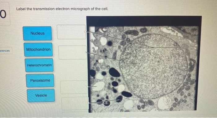
Muscle Lab 19 Figure 19.5 Sarcomere Diagram | Quizlet Start studying Muscle Lab 19 Figure 19.5 Sarcomere. Learn vocabulary, terms, and more with flashcards, games, and other study tools. Solved Label the transmission electron micrograph of the - Chegg Transcribed image text: Label the transmission electron micrograph of the cell. 0 Nucleus rences Mitochondrion Heterochromatin Peroxisome Vesicle ULAR bumit Click and drag each label into the correct category to indicate whether it pertains to the cytoplasm or the plasma membrane. Chapter 14 & 15 Flashcards Flashcards | Quizlet Label the transmission electron micrograph based on the hints provided. Place the following tonsils in order based on their location from superior to inferior. Label the structures in the photomicrograph based on the hints provided. Label This Transmission Electron Micrograph Of A Relaxed ... - Blogger Label this transmission electron micrograph of relaxed sarcomeres by clicking and dragging the labels to the correct location . From science 101 at university high school, tucson. 14… electron micrograph of a relaxed sarcomere in longitudinal section. IMG_2132 - FIGURES Label this transmission electron from
587 Transmission Electron Micrograph Images, Stock Photos ...
IMG_2132.jpg - FIGURES Label this transmission electron micrograph ( 16 ... IMG_2132.jpg - FIGURES Label this transmission electron micrograph ( 16, 000 X ) of a relaxed sarcomere by placing the correct numbers in the spaces Doc Preview Pages 1 Total views 100+ University High School, Tucson Anatomy and Physiology mmederos254 01/26/2019 100% (1) End of preview Upload your study docs or become a member. View full document
Solved] FIGURE 5.5 Transmission electron micrographs of ...
Transmission electron microscopy DNA sequencing - Wikipedia Transmission electron microscopy DNA sequencing is a single-molecule sequencing technology that uses transmission electron microscopy techniques. The method was conceived and developed in the 1960s and 70s, but lost favor when the extent of damage to the sample was recognized. In order for DNA to be clearly visualized under an electron microscope, it must be labeled with heavy atoms.

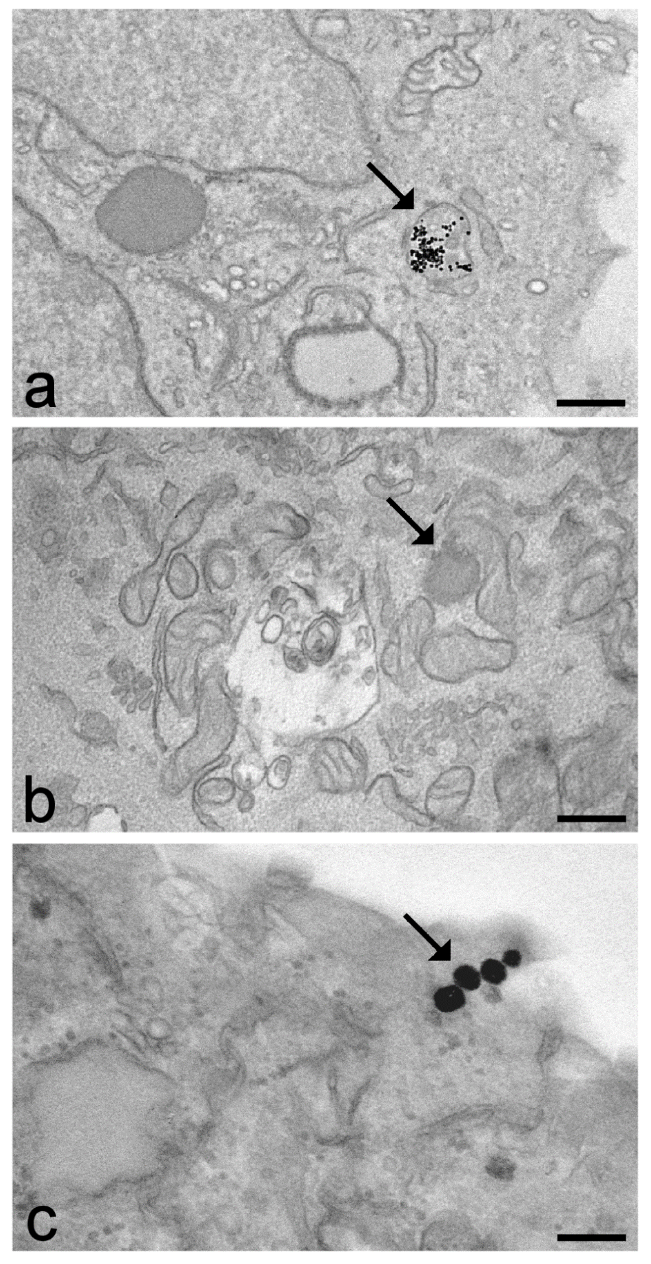
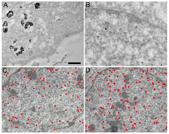
Transmission electron micrographs of the immunogold label for ...
Solved Label the transmission electron micrograph of the - Chegg Label the transmission electron micrograph of the mitochondrion. Matrix granule Mitochondrion Outer membrane Cristae Inner membrane Matrix Reset Zoom Question: Label the transmission electron micrograph of the mitochondrion. Matrix granule Mitochondrion Outer membrane Cristae Inner membrane Matrix Reset Zoom This problem has been solved!
Biology, The Cell, Cell Structure, Eukaryotic Cells | OERTX
Labeling the Cell Flashcards | Quizlet Label the transmission electron micrograph of the nucleus. membrane bound organelles golgi apparatus, mitochondrion, lysosome, peroxisome, rough endoplasmic reticulum nonmembrane bound organelles ribosomes, centrosome, proteasomes cytoskeleton includes microfilaments, intermediate filaments, microtubules Identify the highlighted structures
What is a diagram of a plant and animal cell under an ...
Solved Label the transmission electron micrograph of the | Chegg.com Expert Answer. Answer The label is indicated from TOP to BOTTOM Cil …. Label the transmission electron micrograph of the cilium. Microvillus Axoneme Cilium Dynein arm.

Chapter 14 & 15 Flashcards Flashcards | Quizlet

Solved Mitochondrion Nucleus Vesicle Peroxisome | Chegg.com

Immune electron microscopy - Wikipedia

Muscle Lab 19 Figure 19.5 Sarcomere Diagram | Quizlet

The Transmission Electron Microscope | CCBER

BSC2085L Lab 8 Exercises 12, 13, & 14 Flashcards | Quizlet
Solved Label the transmission electron micrograph of the ...
Solved Label the transmission electron micrograph of the ...

Chapter 14 & 15 Flashcards Flashcards | Quizlet

IMG_2132.jpg - FIGURES Label this transmission electron ...

A&P Unit 2 Exam Flashcards | Quizlet
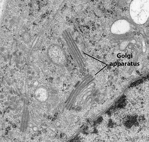
BIOL 230 Lecture Guide - Electron Micrograph of a Golgi Body
IJMS | Free Full-Text | Transmission Electron Microscopy as a ...

lab 12 word.docx - Name: Laboratory Assessment Date: 12 ...

Label electron micrograph of B lymphocyte. - Brainly.com

C2F97E0B-76FD-4F1E-AE0F-47F73CEEAC19 - Laboratory Report 19 ...
f i1w,-
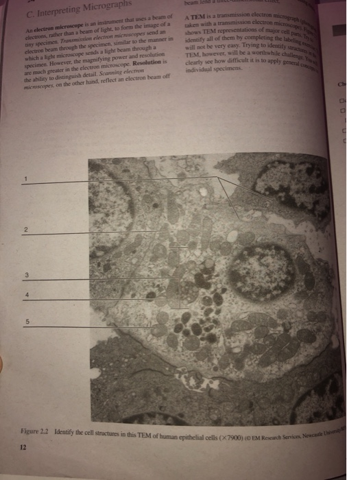
Solved A TEME is a transmission electron micro taken with a ...
Solved Label the transmission electron micrograph of the ...

Solved Label the transmission electron micrograph of the ...
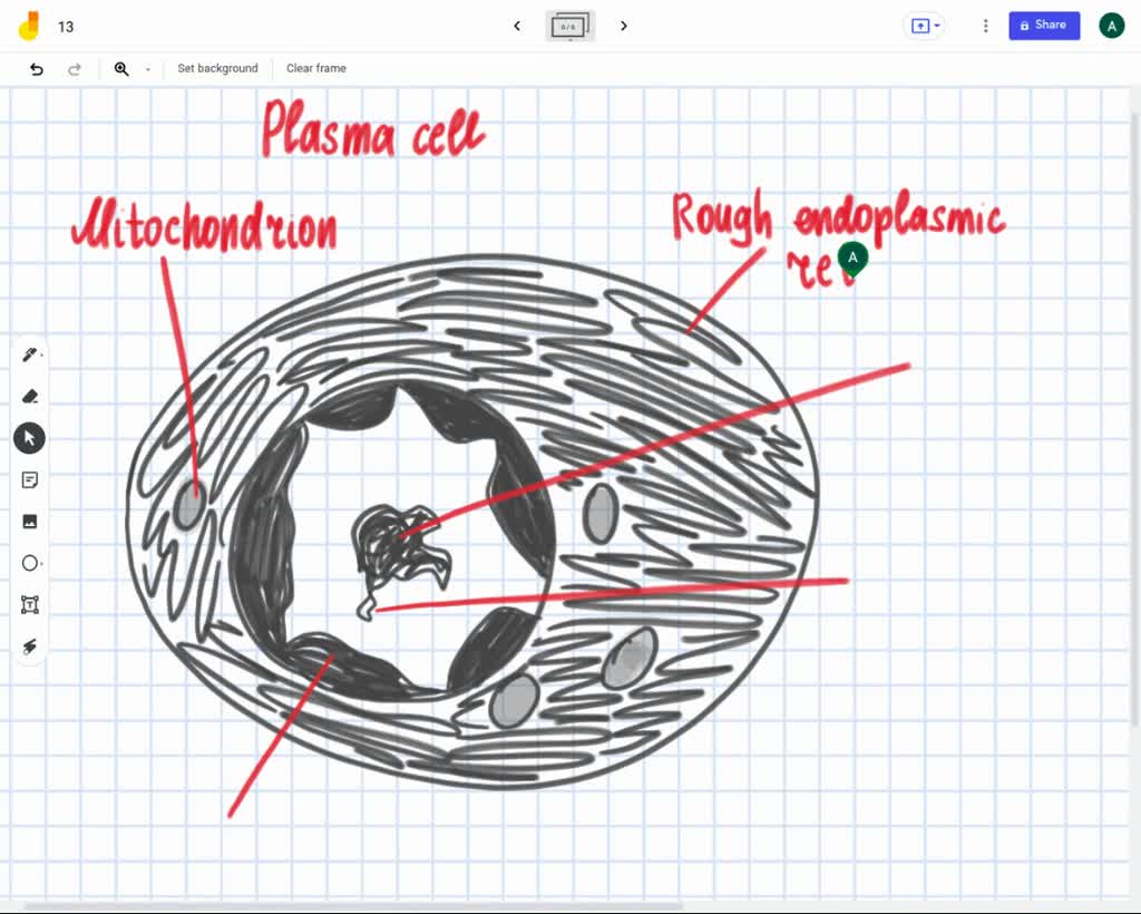
Label the transmission electron ricrograph based on the hints provided, Mitochondnon, Helerochromalin, Plasma cell, Nucleus, Rough endoplasmic Telculn, Nucleolus

What are the labels of the transmission electronic microscope ...
Red blood cell tem hi-res stock photography and images - Alamy

Transmission electron micrograph of a gold-labelled Lowicryl ...
Transmission Electron Micrograph (TEM) showing mitochondria ...
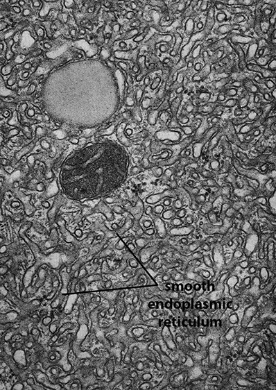
BIOL 230 Lecture Guide - Electron Micrograph of Rough ...

anatomy 10.png - Label the transmission electron micrograph ...

Transmission electron micrograph (TEM) identifying immunogold ...

Overview of the data and gold standard labels for SEM (a) and ...

Transmission electron micrographs of FusMidp-vesicles in ...

Labeling the Cell Flashcards | Quizlet
Live cell immunogold labelling of RNA polymerase II ...

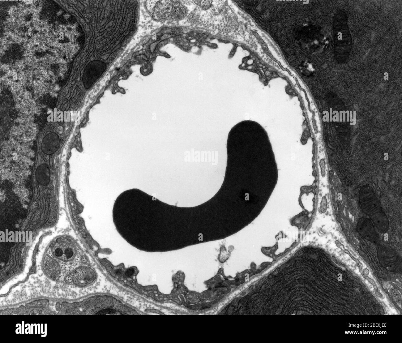
Artery, TEM - Stock Image - C037/6498 - Science Photo Library
Transmission electron microscope (TEM) micrograph showing a ...

Specific, Sensitive, High-Resolution Detection of Protein ...
A and B) Electron micrograph of a cell labeled for/5-tubulin ...

Transmission electron micrograph of mature MRCs with anti-Na ...


Komentar
Posting Komentar